Signal Processing in the DS ganglion cell dendritic tree
Robert G. Smith
We are currently studying signal processing performed by the ganglion cell of the mammalian retina. A ganglion cell is responsible for transmitting the visual signal from the retina to the brain. It must generate a series of action potentials (spikes) in response to a light flash. Different ganglion cells filter and convert the signal into spikes each using a different code.
Sodium and potassium channels are necessary in the soma and axon of most neurons for generating spikes. A spike is generated in the initial segment of the axon in response to depolarization of the soma by synaptic inputs in the dendritic tree.
However, sodium and potassium channels are also necessary in the dendritic tree. The reason, discovered by Fohlmeister & Miller (1997a,b) is that a spike initiated in the soma propagates into the dendritic tree and charges up the capacitance in the dendritic membrane. If not discharged by an active spike, the charge in the dendritic membrane flows back to the soma and causes the next spike to initiate too soon, increasing the minimum spike rate.
Therefore, to allow the neuron to spike slowly, sodium and potassium channels are necessary in the dendrites. The dendrites can then actively propagate spikes and discharge their membrane capacitance after each spike. Sodium channels are known to exist on the dendritic tree of alpha ganglion cells and also direction-selective (DS) ganglion cells, and are thought to exist in the dendritic trees of most ganglion cell types.
The DS ganglion cell is unusual in that it initiates spikes in its dendritic tree. Whether a spike is initiated or not is a decision made by the dendrite, and this represents nonlinear processing that amplifies the direction-selective signal (Oesch et al, 2005). The spikes propagate to the soma and then back-propagate to the remainder of the dendritic tree.
We are studying how the voltage-gated channels in the dendritic membrane process the synaptic signals. If excitatory synaptic inputs are balanced by inhibitory inputs, no spikes will occur. However, if excitatory post-synaptc potentials (EPSPs) are momentarily synchronized betwen different synaptic sites along a dendrite, the resulting transient depolarization may be enough to initiate a spike. Once a spike is initiated, it may propagate forward to the soma and axon. We found that even very strong inhibition on the path to the soma does not block a spike from propagating. Mike Schachter developed the model and made a movie of the spike propagation
However, if another dendrite has just initiated a spike, the sodium channels on the path to the soma may be inactivated, momentarily preventing any other spikes from propagating back to the soma.
We are continuing to study the DSGC and its presynaptic circuitry

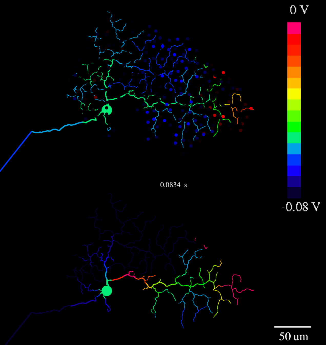
Figure 1. Spike initiation in the DS ganglion cell. Top image shows On-layer of bistratified dendritic tree. Dendrites are colored according to the membrane voltage (scale bar on right). Most of the dendritic tree is hyperpolarized (blue). Excitatory synaptic inputs (red dots) at rightmost portion of dendritic tree are generating a spike (rightmost dendrite, red). A previous spike leaves the path to the soma partially depolarized (green). Bottom image shows inactivation of same dendritic tree. The new spike can propagate part way along the path to the soma because it has mostly recovered from inactivation. However, one segment of the proximal dendrite is still partially inactivated.
|

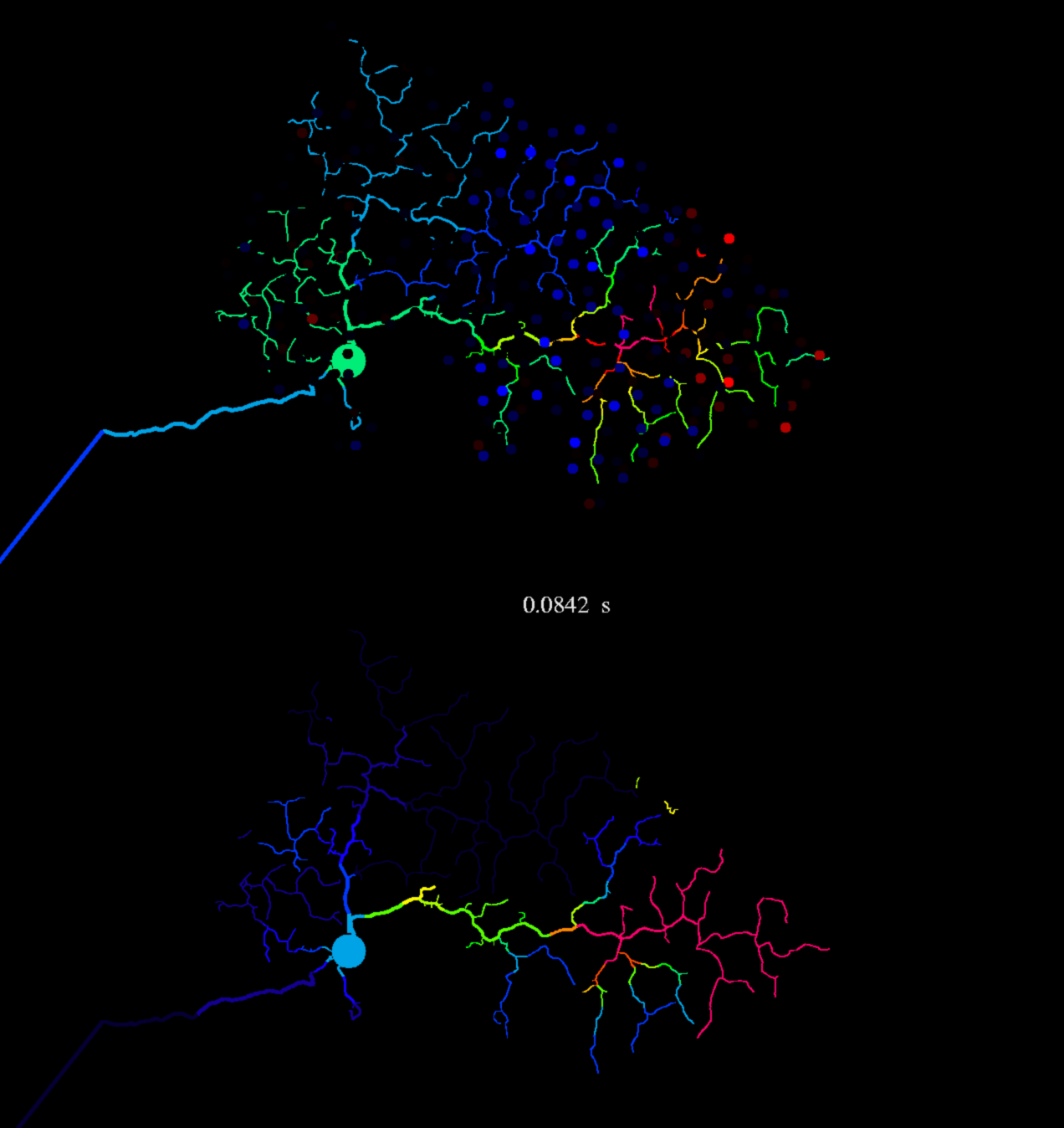
Figure 2. Spike propagation. Top image (color=voltage), 0.8 msec later, spike has propagated ~50 um towards the soma, Bottom image (color=inactivation) Region behind spike is inactivated (red). Region ahead of spike is still partly inactivated (green, yellow). Synaptic inhibition (blue dots) does not block actively propagating spike because sodium conductance is greater than inhibitory conductance.
|

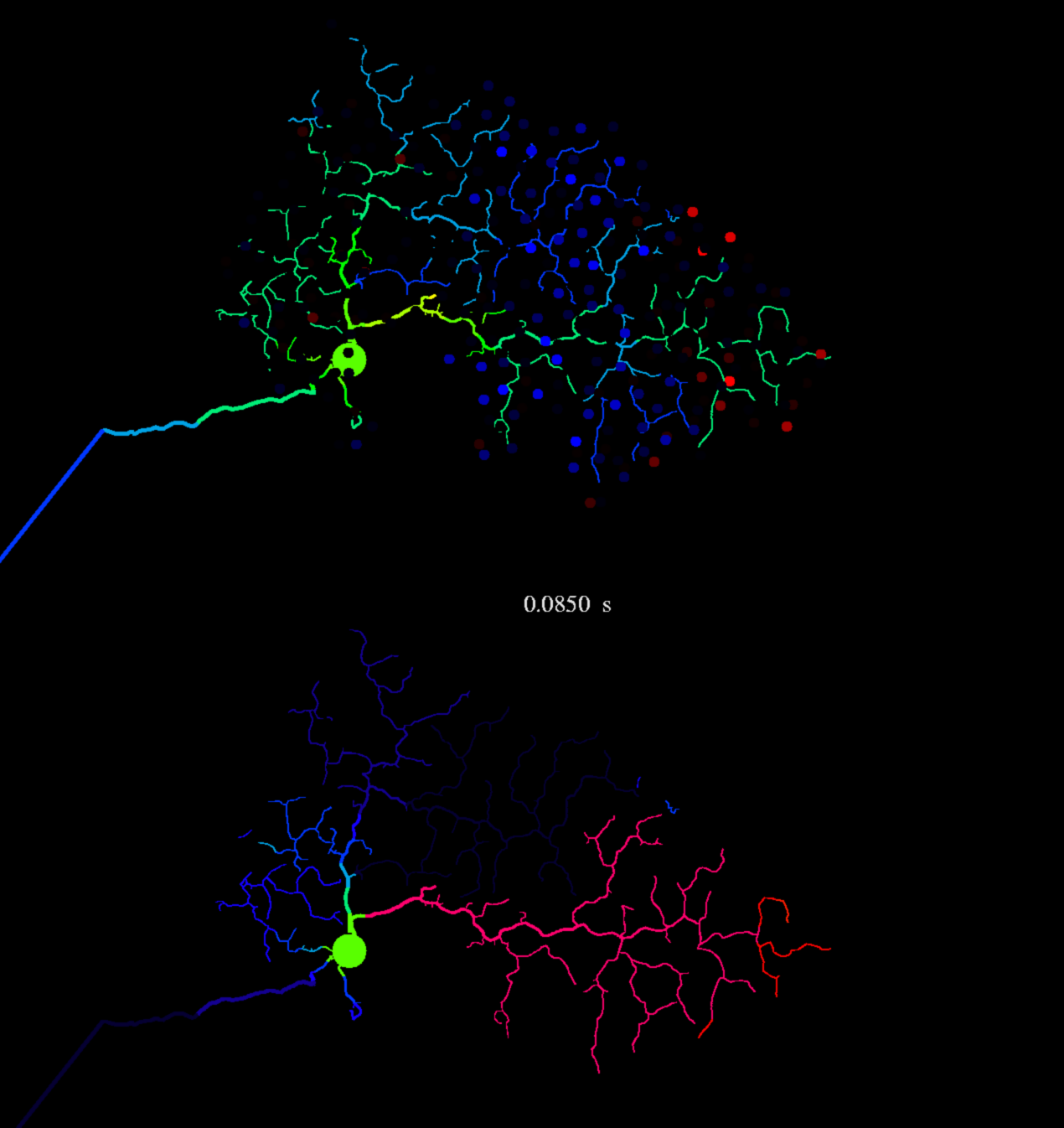
Figure 3. Spike block. Top image, after another 0.8 msec, the propagating spike has almost dissipated (yellow) before it reaches soma. Bottom image, all of dendrite is inactivated from the depolarization in the spike (red). |
|
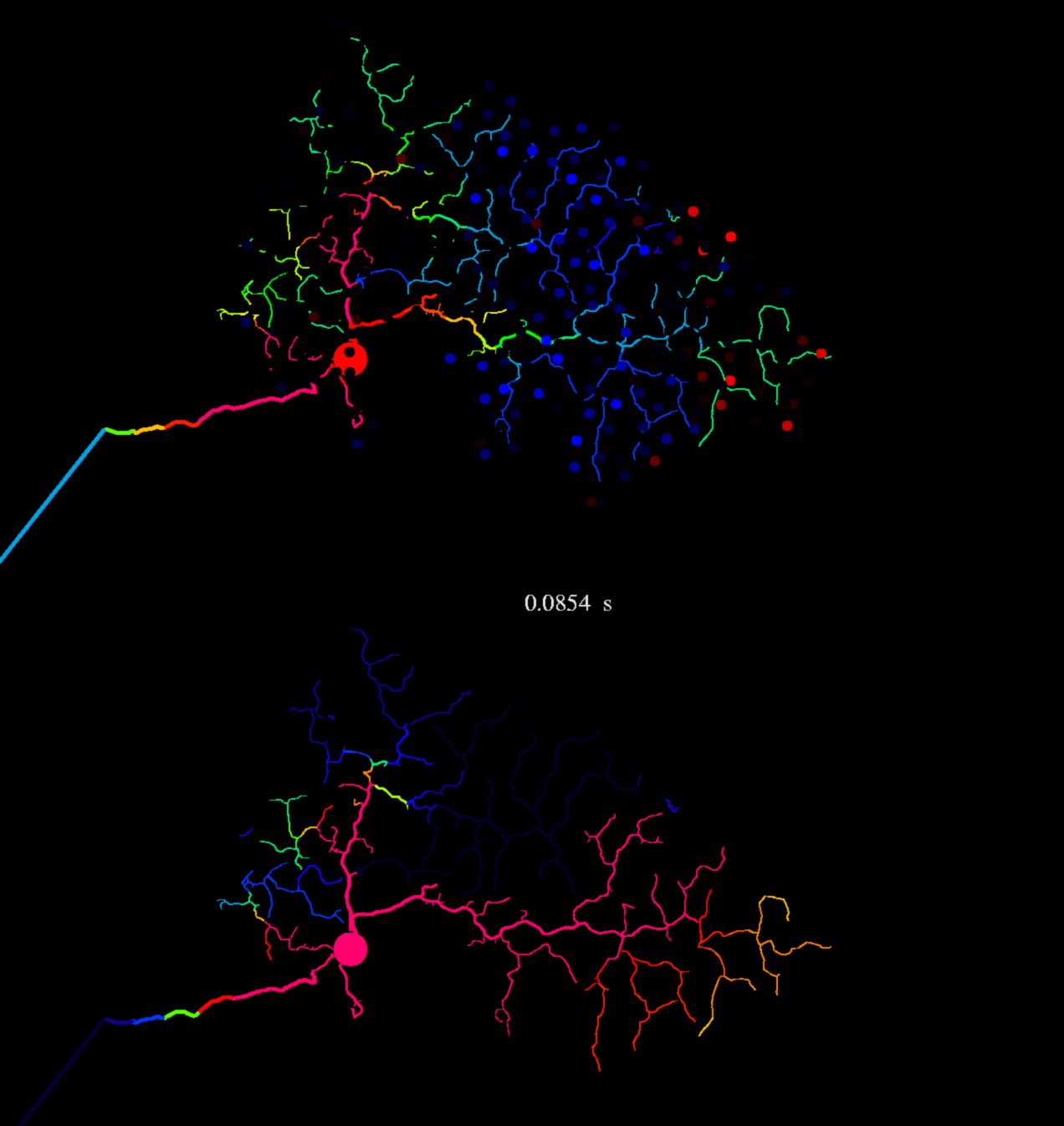
Figure 4. Spike back-propagation. Top image, after another 0.4 msec, spike recovers and propagates to soma, then propagates to the other dendrites. Bottom image, dendrite where spike originated is inactivated. Rightmost dendritic tip is recovering from inactivation, so it may be able to initiate the next spike.
|
